
What is Vein Mapping?
Have you ever felt aching, itching, heaviness, burning sensation, fatigue, restlessness, throbbing, or cramping in your body? If yes then it means that these are the symptoms of vein disease. The next step will be Vein mapping.
It is also called vascular ultrasound is used in the diagnosis and treatment of vein disease. It is an ultrasound test, done to analyze all the veins in your legs and /or arms. It creates a “map” of where your abnormal (varicose) veins are located. Varicose veins are painful veins on or near the surface of the legs. They are usually caused by venous insufficiency, also known as venous reflux.
Vein Mapping Procedure:
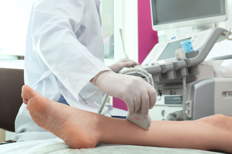
How is vein mapping done? Ultrasounds transmit high-frequency sound waves to the varicose veins in the body. Sound waves are recorded and transformed into images. These images are then used to create a map of the arteries and veins and any blockages in them. Sound waves can also track the speed of blood cells flowing within blood vessels and are displayed on the screen. With this information, your doctor can diagnose vein disease and determine the best treatment plan available.
Vein mapping ultrasound protocol:
Ultrasound vein mapping, also known as vascular ultrasound, is a non-invasive diagnostic procedure that does not require the use of needles, dyes, radiation or anaesthesia.
Ultrasound tests send high-frequency sound waves through the tissues of your body. An ultrasound machine records sound echoes and convert them into videos or images of body tissues and fluids. During the procedure, a vein mapping machine transmits sound waves through a section of blood vessels in your body. These sound waves reflect the blood cells moving inside your blood vessels. The sound waves can also record the speed of your blood flow and are displayed on a computer screen.
Vein mapping for bypass surgery:
If your consultant has decided that you need bypass surgery (CABG) for a blocked artery in your heart or leg, they may refer you for a vascular ultrasound called vein mapping for CABG.
During bypass surgery, the surgeon will need to take a vein, from another part of your body usually from your leg to use as a replacement artery to redirect blood flow around the blocked arteries. Before they do this your provider may use vascular mapping to find the correct blood vessel to use. During the evaluation, a vascular scientist will apply some cold gel to your leg with an ultrasound scanner, which will image the superficial veins in your leg. A series of vein measurements will be taken, and any problems such as blocked areas of the vein will also be checked and reported. Depending on the type of bypass you need and your doctor’s needs, we may need to draw a line on your leg to guide the surgeon through the procedure.
Vein Mapping lower and upper extremities:
What is lower extremity vein mapping? Vascular mapping is the process of identifying and measuring veins in the upper or lower extremities. The lower extremity is done for coronary artery bypass grafting (CABG).
By measuring the diameter of a particular vein and checking blood flow, a physician can determine whether a patient is suffering from a condition known as venous insufficiency. Another use of these tests is to determine if a patient has a blockage or thrombus in the vasculature. To assess and size the venous system for thrombosis and to determine the suitability of the veins for possible arterial bypass, or for the creation of dialysis access grafts, fistulas, and carotid artery bypass grafts.
The upper extremity is performed to evaluate the veins in your arms to determine the diameter, length and size for use during dialysis surgery or arterial bypass surgery in your arm or leg. The upper extremity arterial system and The central venous system are assessed for patency before dialysis access is established. The anatomy of the veins and arteries of the UE is essentially the same as that of the lower extremities, although some structures are more prominent than others and the nomenclature differs.
The veins of the upper extremity are subject to less pressure than the veins of the lower extremity. In the standing position, the hydrostatic pressure due to the fluid column from the heart to the feet is in the range of 90 mmHg (depending on length). The lower veins are thicker than the upper veins to resist these higher pressures. For this reason, they have better baseline patency than UE veins when used for arterial bypass. Lower extremity veins also have more valves, which facilitate the pumping action of muscle contractions.
Vein mapping for kidney dialysis:
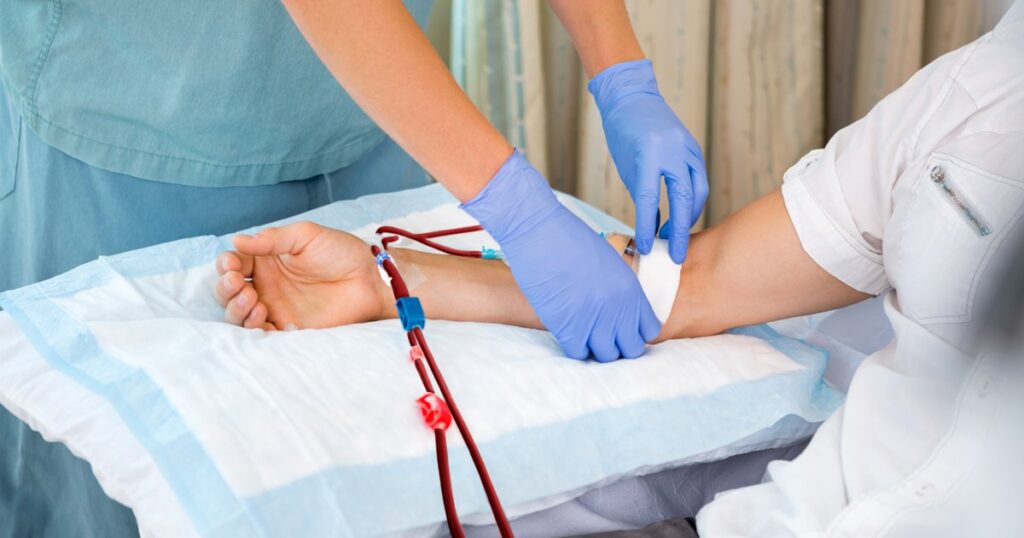
Your kidney doctor has decided that you have advanced kidney disease and will need dialysis sometime in the future. If you have chronic kidney disease (CKD), you may need dialysis. the preferred dialyses are peritoneal dialysis and hemodialysis. If your doctor suggests hemodialysis, an artificial kidney, or dialyzer, will work to remove waste and fluid from your blood. Starting hemodialysis requires creating vascular access, also called hemodialysis access, to take your blood to the kidney.For this, vein mapping is done to help decide which arm and which artery and vein in the arm should be used to create a fistula or graft.
The choices of hemodialysis access are
- Arteriovenous fistulas (AFVs)
- Arteriovenous grafts (AVGs)
- Central venous catheters(CVC).
Hemodialysis treatments are scheduled three times per week at a dialysis clinic or home dialysis may be an option for some people.
To create an arteriovenous fistula, a vascular specialist will connect a vein to an artery in your non-dominant arm.
If for some reason the blood vessels in the arms are found to be of insufficient size to create an arteriovenous fistula, a graft may be used instead. If this happens, a soft, flexible biocompatible tube will be placed under the skin, with one end connected to the chosen artery, and the other to the chosen vein, thus connecting the vein and artery.
A central venous catheter (CVC) is another type of vascular access. With a CVC, a tube is placed inside a central vein in your chest with its tip directly into the upper right chamber of your heart. The doctor numbs the area of the skin for insertion and may use imaging to guide the correct placement of the catheter.
Vein mapping for venous insufficiency:
Vein Mapping is done to measure the diameter of a particular vein and examine blood flow, to determine if a patient is suffering from venous insufficiency. Chronic venous insufficiency (CVI) is the set of symptoms and signs that affects the lower extremities due to poor valvular function of the venous systems. CVI happens when your legs do not allow blood to flow back to your heart. Blood may pool in your legs, known as venous reflux disease or venous insufficiency/vein disease. Every venous insufficiency is the consequence of a valuation, which results primarily in varicose veins. Venous insufficiency often manifests as leg pain, heaviness in the legs, leg discolouration or varicose veins. Other symptoms of chronic venous insufficiency may include swelling in your legs or ankles, itching, painful legs, pain while walking that stops when you rest, open sores on the legs (leg ulcers), leg cramps or spasms, or an uneasy urge to move your legs (restless leg syndrome). Left untreated, venous insufficiency can cause more serious difficulties like ulcers, bleeding, and a life-threatening condition called Deep Vein Thrombosis (DVT).
FAQ
Is vein mapping painful?
No. As this procedure uses ultrasound, it is a non-invasive and painless procedure. There is no need for needles, dyes, anaesthesia and incisors. This imaging test has no harmful side effects because ultrasound does not use radiation and has no harmful effects on the human body.
How long is vascular mapping?
A complete vein mapping study may take up to 30-90 minutes to complete. The physician will need access to your legs from the groin to the ankle or your arms from the neck to the wrist.
What is vein mapping used for?
It is a risk-free and non-invasive procedure that uses ultrasound technology to see if the veins are suitable for the desired type of surgical procedure (bypass) including open-heart surgery, bypass grafts in the legs or the creation of a fistula for dialysis. It is a technique used to determine the patient’s anatomy, course, and quality. It can be used to guide certain medical procedures or diagnose certain health conditions.
How much does vein mapping cost?
The cost of saphenous vein mapping varies from case to case. Your doctor will determine if the procedure is required and provide you with a cost estimate. This procedure typically costs $300-$500 per session. So the total cost of vascular mapping can range from $300 to $2000.
What is vein mapping for a fistula?
It is used in the surgical placement of an arteriovenous fistula (AV fistula), which is created by connecting an artery to a vein using your vessel. It helps ensure that the veins are of sufficient size and flow to support the fistula. The surgeon usually places the AV fistula in the arm or upper arm. An AV fistula causes extra pressure and extra bleeding in the vein, making it larger and stronger. A large vein provides easy and reliable access to blood vessels. Without this vascular access, regular hemodialysis sessions would not be possible. It takes about three to six months for a fistula to mature enough to be used, so vein mapping and fistula surgery need to be done well before starting dialysis treatment.